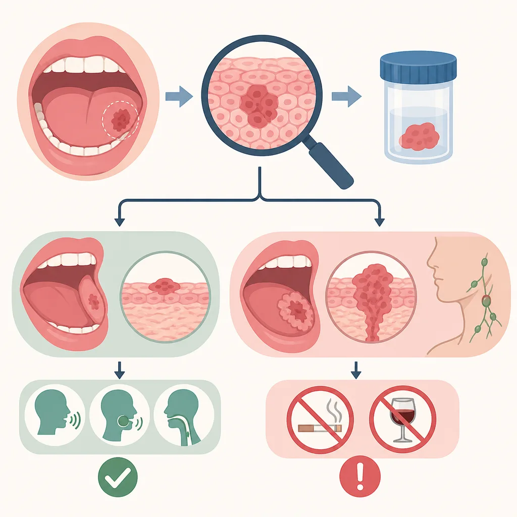
Oral Cancer Symptoms and Diagnosis: The Warning of a Persistent Mouth Sore
Most mouth sores are not cancer, but an ulcer that does not heal for at least 2 weeks, red or white patches, lumps, or unexplained bleeding are warning signs that require medical attention. Oral cancer is confirmed through a biopsy, and earlier detection is more likely to reduce the extent of treatment and loss of function.
- If an ulcer or patch in the mouth persists for at least 2 weeks, its cause should be evaluated by a dentist or an otolaryngologist.
- Smoking and excessive alcohol consumption are major risk factors for oral cancer, and the risk increases further when both factors are present.
- Oral cancer cannot be definitively diagnosed based on appearance or pain alone; a biopsy of the suspicious area is required.
- The extent of surgery and method of reconstruction are determined by the tumor's location, size, depth of invasion, lymph node metastasis, and the patient's functional status.
- Early detection is important not only for improving the chances of survival but also for preserving speech, chewing, and swallowing functions.
Most mouth sores are not oral cancer. Common aphthous ulcers or traumatic wounds usually improve on their own, but lesions that do not heal within a certain period or continue to grow require examination. In particular, oral cancer may not cause obvious pain in its early stages, so it is important to monitor the duration of symptoms and pattern of changes rather than their severity.
This article is not intended for self-diagnosis. If you have a persistent lesion, bleeding, difficulty swallowing, or a lump in the neck, you should be evaluated by a healthcare professional.
What Is Oral Cancer?
Oral cancer refers to malignant tumors that develop in the lips and tissues inside the mouth. Most oral cancers are squamous cell carcinomas, which begin in the squamous cells that form the mucosal lining.
Main Areas Included in the Oral Cavity
- Inner lips and buccal mucosa
- Front approximately two-thirds of the tongue
- Floor of the mouth beneath the tongue
- Upper and lower gums and alveolar mucosa
- Hard palate
- Retromolar trigone behind the wisdom teeth
The base of the tongue, tonsils, and soft palate are generally part of the oropharynx, not the oral cavity. Therefore, cancers of the tonsils and base of the tongue are classified as oropharyngeal cancers rather than oral cancers. Salivary gland cancers may also be found inside the mouth, depending on where they arise, but they are treated as a separate group of tumors.
Differences Between Mouth Ulcers and Lesions Suspicious for Oral Cancer
It is impossible to distinguish benign mouth ulcers from cancer perfectly based on appearance alone. The following table provides reference criteria for deciding whether medical attention is needed; a definitive diagnosis is made through a professional examination and biopsy.
| Category | Common Mouth Ulcers and Traumatic Wounds | Suspicious Findings Requiring Medical Attention |
|---|---|---|
| Duration | Usually improve within 1–2 weeks | Do not heal for at least 2 weeks or persist for up to 3 weeks |
| Changes | Gradually become smaller and less painful | Become larger or deeper, with hardening of the surrounding area |
| Surface and color | Round ulcers with red borders are common | Persistent white or red patches, or lesions containing both colors |
| Texture | May be temporarily painful | A firm lump or tissue hardening can be felt |
| Accompanying symptoms | Local pain when irritated | May be accompanied by bleeding, reduced sensation, difficulty swallowing or speaking, or a neck lump |
| Course after removing the cause | Improves after eliminating irritation such as a sharp tooth | Does not heal even after the cause is removed |
The commonly cited “2-week rule” is not a rule that confirms cancer but a safety threshold to avoid delaying an examination. If a wound persists for at least 2 weeks, make an appointment, and if it has not healed for at least 3 weeks, it should be evaluated. If bleeding does not stop or problems with breathing or swallowing occur, seek prompt medical attention regardless of how long the symptoms have been present.
Warning Signs That Should Not Be Missed
The following symptoms do not all mean cancer, but they should be checked if they persist or worsen.
- An ulcer on the lip or inside the mouth that does not heal for at least 2 weeks
- A white patch that cannot be rubbed off or a vivid red patch
- A firm lump felt inside the mouth, on the tongue, or in the gums
- Recurrent bleeding without an apparent reason
- Reduced sensation or numbness of the tongue or lips
- Changes in chewing, swallowing, speaking, or tongue movement
- Persistent pain that radiates to one ear
- Dentures that previously fit well suddenly becoming uncomfortable
- Unexplained loose teeth or an extraction site that does not heal
- A newly felt lump under the jaw or in the neck
- Unexplained weight loss
Bad breath can result from many causes, including oral hygiene, periodontal disease, and tonsil conditions, so it cannot by itself be used to identify oral cancer. Conversely, early oral cancer may be painless.
Are All White and Red Patches Cancer?
No. Leukoplakia is a clinical term describing a white lesion that does not disappear when wiped, while erythroplakia refers to a distinct red lesion. Other causes, such as infection, friction, and inflammation, are also possible.
However, some lesions may contain epithelial dysplasia, which is a precancerous change, or cancer. Healthcare professionals assess the lesion’s location, size, texture, and risk factors and perform a biopsy when necessary. A lesion should not be judged benign or malignant based on its visible color alone.
Major Risk Factors
1. Tobacco and Other Tobacco Products
Tobacco products, including cigarettes, cigars, pipes, and smokeless tobacco, are major risk factors for oral cancer. This is because the mucous membranes of the mouth and pharynx are repeatedly exposed to carcinogens in tobacco smoke and products.
2. Excessive Alcohol Consumption
Excessive alcohol consumption increases the risk of oral cancer. When smoking and drinking continue together, the risk rises more sharply than when only one is present. Total alcohol intake and drinking patterns are more important than the type of alcoholic beverage.
3. HPV Infection
High-risk human papillomavirus, particularly HPV 16, is associated with some head and neck cancers. However, the association with HPV is especially clear in oropharyngeal cancers arising in the base of the tongue and tonsils, which should be distinguished from typical oral cancers that develop in the front of the mouth.
4. Betel Quid and Chewing Mixtures
The habit of chewing betel quid or areca nut and mixtures containing them increases the risk of oral cancer. This is a particularly important risk factor in some regions of Asia and the Pacific.
5. Ultraviolet Exposure of the Lips
Long-term ultraviolet exposure through outdoor work or activities may be associated with the risk of lip cancer, particularly cancer of the lower lip.
6. Age and Medical History
Oral cancer is more common among middle-aged and older adults, but it can also occur in younger people. Those with a history of head and neck cancer or impaired immune function should follow the surveillance schedule established by their healthcare professionals.
Is Chronic Irritation a Direct Cause?
Poorly fitting dentures, sharp teeth, and repeatedly biting the cheek or tongue can cause ulcers and inflammation. However, there is insufficient evidence to conclude that this type of physical irritation alone causes oral cancer. What is important is to undergo an evaluation, including a biopsy, if the lesion does not heal even after the source of irritation has been corrected.
How Is Oral Cancer Diagnosed?
1. Medical History and Examination of the Mouth and Neck
Healthcare professionals ask when the symptoms began and assess smoking and alcohol use, weight changes, pain, and swallowing problems. Using a light and mirror, they examine the lips, cheeks, gums, sides and underside of the tongue, floor of the mouth, and palate, and feel for lumps inside the mouth and in the neck.
2. Biopsy
A definitive cancer diagnosis requires a biopsy, in which suspicious tissue is removed and examined under a microscope. Tests that collect cells with a brush or adjunctive tests using light may assist in evaluation, but they generally cannot replace a biopsy of a suspicious lesion.
3. Imaging and Staging
Once cancer is confirmed, tests such as CT, MRI, ultrasound, and PET-CT may be performed as appropriate to assess tumor depth, invasion of surrounding tissues, cervical lymph nodes, and distant metastasis. The stage is determined by considering the tumor’s size and depth of invasion, lymph node metastasis, and distant metastasis.
How Is Treatment Determined?
Treatment is planned jointly by specialists in head and neck surgery, radiation oncology, medical oncology, dentistry, reconstructive surgery, and rehabilitation, taking into account the tumor’s location and stage, whether it can be removed, pathology findings, the patient’s overall health, and the possibility of preserving function.
| Treatment | Use and Purpose |
|---|---|
| Surgery | Removes the local tumor and, when necessary, cervical lymph nodes. It is commonly used as a key treatment for early oral cancer. |
| Radiation therapy | May be used alone or after surgery to reduce the risk of recurrence. |
| Chemotherapy | May be combined with radiation therapy for advanced cancer or used for recurrent or metastatic disease. |
| Targeted therapy | In certain circumstances, drugs that block cell growth signals may be used. |
| Immunotherapy | Drugs that activate the immune response are used for some recurrent or metastatic head and neck squamous cell carcinomas. |
During surgery, the tumor is removed with a margin of normal tissue, but the same 1–1.5cm margin is not applied to every patient. The required extent varies according to the tumor’s location, depth of invasion, relationship to critical structures, and the results of intraoperative and postoperative pathology examinations.
Reconstructive Surgery After Resection
Small lesions may recover through simple suturing or natural healing. Larger defects may require reconstruction to preserve speech, chewing, swallowing, and facial appearance.
Common Reconstructive Methods
- Radial forearm free flap: Uses thin, flexible skin and blood vessels from the inner forearm to reconstruct soft tissues such as the tongue or floor of the mouth.
- Anterolateral thigh flap: Uses skin and soft tissue from the thigh and is employed to fill relatively large defects.
- Fibula free flap: Transfers part of the fibula, along with blood vessels and soft tissue, to reconstruct the lower jaw.
- Local or pedicled flap: Moves nearby tissue or tissue with its blood supply still connected, depending on the defect’s location and size.
In free flap surgery, a microscope is used to connect the small blood vessels of the transferred tissue to blood vessels in the neck. Technologies such as CT-based virtual surgical planning, cutting guides, and patient-specific metal plates may be used when necessary, but they are not required for every patient.
Rehabilitation and Long-Term Care After Treatment
Oral cancer treatment does not end with removal of the tumor. Surgery and radiation therapy can affect speech, swallowing, saliva production, taste, jaw movement, and dental health.
Main Areas of Rehabilitation
- Speech and communication training through speech therapy
- Swallowing assessment and training in safe food intake
- Nutritional assessment and adjustment of food consistency
- Prevention and exercise for restricted mouth opening
- Dental and gum care and prosthetic rehabilitation
- Prevention of dry mouth and tooth decay after radiation therapy
- Psychosocial support for pain, anxiety, depression, and changes in appearance
- Support for stopping smoking and alcohol consumption
When swallowing is impaired, aspiration may occur when food or saliva enters the airway. A professional swallowing assessment is needed if there is coughing during meals, a wet-sounding voice, recurrent pneumonia, or a marked increase in mealtime duration.
How to Interpret Early Detection and Survival Rates
Oral cancer survival rates should not be generalized into a single number. Outcomes vary greatly depending on the site of origin, stage at diagnosis, lymph node metastasis, biological characteristics of the tumor, treatment options, and the patient’s overall health.
In general, oral cancer detected at a localized stage has a better prognosis than cancer that has spread to cervical lymph nodes or other organs. Treating a lesion while it is small also increases the likelihood of reducing the extent of resection, the need for reconstruction, and functional loss. However, even early cancers may require extensive treatment depending on their location and depth of invasion, while advanced cancers may be treated or controlled for long periods through a combination of surgery, radiation, and drug therapy. Treatment should therefore not be abandoned based on stage alone.
Prevention and Everyday Care
- Do not use tobacco products, and obtain smoking cessation support if you currently use them.
- Avoid alcohol or reduce consumption, and avoid the combined habit of smoking and drinking.
- Do not chew betel quid or areca nut products.
- Use lip sunscreen and a wide-brimmed hat during outdoor activities.
- Have sharp teeth, broken dental restorations, and poorly fitting dentures corrected by a dentist.
- During regular dental visits, have not only the teeth but also the tongue, floor of the mouth, and buccal mucosa examined.
- Consult a healthcare professional and consider HPV vaccination according to the recommended age and schedule.
- Record new lesions with photographs and dates, but do not delay medical attention because of self-monitoring.
Looking inside the mouth with a mirror can help people notice changes, but a normal appearance cannot rule out cancer. This is because areas under the tongue or deep in the neck are difficult to inspect on your own.
When Should You Seek Medical Attention?
Evaluation by oral medicine, oral and maxillofacial surgery, or otolaryngology is recommended in the following situations.
- A wound, patch, or lump inside the mouth persists for at least 2 weeks
- The lesion grows rapidly or the surrounding area becomes firm
- There is recurrent bleeding, numbness, or a neck lump
- Difficulty swallowing or speaking persists
- There is no apparent explanation for loose teeth or delayed healing of an extraction site
If breathing is difficult, even saliva is hard to swallow, or heavy bleeding does not stop, seek emergency medical care immediately.
FAQ
Should all mouth sores be suspected of being oral cancer?
No. Benign causes such as aphthous ulcers, wounds from biting the cheek or tongue, and trauma from sharp teeth are far more common. However, if a sore does not heal for at least 2 weeks or becomes larger and harder, you should seek medical attention.
Is oral cancer always painful?
No. Early oral cancer may be painless or cause only mild discomfort. More important warning signs than pain may include how long the lesion remains unhealed, changes in size, hardening, bleeding, and reduced sensation.
Is a mouth ulcer that persists for at least 2 weeks always cancer?
It is not necessarily cancer. There are many possible causes, including infection, repeated irritation, immune disorders, and medications. However, because it has persisted beyond the time a typical wound would take to heal, it is safer to have the cause evaluated by a dentist or an otolaryngologist.
Can oral cancer be diagnosed with a blood test?
Oral cancer cannot be definitively diagnosed with routine blood tests alone. The key to a definitive diagnosis is a biopsy, in which the suspicious lesion is examined directly and a tissue sample is collected and examined under a microscope.
Are all white patches in the mouth precancerous lesions?
No. White patches can result from various causes, including friction, fungal infections, and inflammation. Persistent white or red patches that do not disappear when wiped may require evaluation because epithelial dysplasia or cancer may be found in some cases.
Can nonsmokers also develop oral cancer?
Yes. Smoking is an important risk factor, but oral cancer can also occur in nonsmokers. Various factors are associated with it, including alcohol consumption, betel nut use, ultraviolet exposure, age, and immune status, and some patients have no clear risk factors.
Is HPV the main cause of all oral cancers?
No. High-risk HPV is strongly associated with oropharyngeal cancer arising in the tonsils and base of the tongue, but smoking and alcohol consumption are often more important risk factors for typical oral cancers in the front part of the mouth.
Does oral cancer surgery always involve extensive removal of the tongue or jawbone?
No. The extent of removal varies depending on the cancer's location, size, depth of invasion, and whether it has invaded surrounding tissues. Small, early-stage lesions can be removed with limited surgery, but advanced lesions may require more extensive surgery and reconstruction to preserve function and ensure clear surgical margins.
Can oral cancer be definitively diagnosed through a dental examination alone?
Dental examinations can help detect suspicious lesions early, but a definitive diagnosis cannot be made by visual examination alone. If suspicious findings are present, a specialist examination and biopsy are necessary.
Can speech and eating ability recover after oral cancer treatment?
The degree of recovery varies depending on factors such as the extent of removal and whether radiation therapy was administered. Function can be improved through reconstructive surgery, speech and swallowing rehabilitation, nutritional care, and dental treatment, and it is important to begin systematic rehabilitation early.
Sources
- National Institute of Dental and Craniofacial Research: Oral Cancer
- National Cancer Institute: Lip and Oral Cavity Cancer Treatment
- National Cancer Institute: Head and Neck Cancers Fact Sheet
- NHS: Mouth Cancer
- American Cancer Society: Oral Cavity and Oropharyngeal Cancer
- World Health Organization: Oral Health Fact Sheet
Images